Scientists at the Moscow Institute of Physics and Technology (MIPT) have synthesized a molecule that may be able to fight chemoresistant ovarian cancer, according to a study recently published in the the European Journal of Medicinal Chemistry.
The molecule, which was described in the study, “Synthesis and anti-mitotic activity of 6,7-dihydro-4H-isothiazolo[4,5-b]pyridin-5-ones: In vivo and cell-based studies,” may be helpful in the treatment of ovarian cancer patients whose disease recurred following chemotherapy.
The researchers developed 37 different compounds belonging to a class known as aminoisothiazoles which are used in a wide range of pharmacological and biological applications, and assessed their anti-tumor properties.
“We made the decision to experiment with aminoisothiazoles because compounds of this class exhibit a diverse range of pharmacological and biological activities. This led us to expect that with the appropriate functional groups, they might act as anticancer agents,” Alexander Kiselyov said in a press release.
The compounds were developed through a straightforward approach that involved six steps, and which used standard, easily obtainable reagents. Then the researchers tested each compound on human cancer cell lines from breast adenocarcinoma, melanoma, ovarian, and lung tumors, as well as sea urchin embryos.
Of the 37 compounds, the team found that 12 could reduce the proliferation rate of cancer cells, with some preventing their proliferation altogether, which led to cancer cell death. One compound in particular was found to exert an effect in chemoresistant ovarian tumor cells.
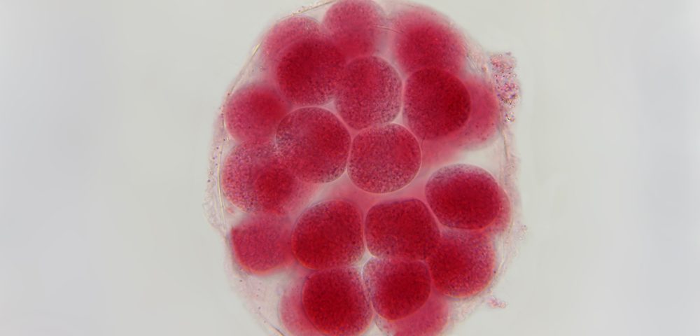
The researchers believed that the anti-cancer properties of these compounds was due to their effects on tubulin, a component from the cell cytoskeleton involved in cell division. So they tested the newly synthesized compounds in sea urchin embryos. Previous studies have shown that sea urchin embryos will rapidly spin when anti-tubulin agents interrupt cell division, and their activity is easily detected by light microscopy.
Researchers can also study the embryos to look at other important factors in determining a molecule’s anti-cancer properties, including specific anti-proliferation effects, solubility, overall toxicity, and biomembrane permeability. Also making it an ideal test cell, the sea urchin embryo cell’s rate of mitosis is 40 minutes, compared to 24 hours for a human cancer cell, and is more sensitive to the agents used in the study.
Importantly, the molecule that could affect the ovarian cancer chemoresistant cells was found to be the most potent anti-cancer compound in the sea urchin studies.
The researchers concluded that the newly developed molecule displays anti-tubulin activity and can destroy the chemoresistant ovarian carcinoma cells. They now plan on using crystallography data and structure modeling to further investigate microtubule degradation and to identify tubulin binding sites.

