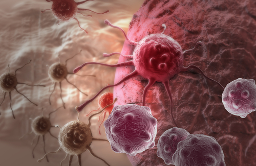
A lab-grown, three-dimensional (3D) ovarian tumor model will give researchers a new way to study tumor growth in ovarian cancer and to test how tumors might respond to treatments, scientists say.
The proof-of-concept study, “Peptide-protein coassembling matrices as a biomimetic 3D model of ovarian cancer,” was published in the journal Science Advances.
Preclinical studies of potential cancer therapies rely largely on two-dimensional tissue cultures — cells grown in lab dishes — and animal models of the type of disease being treated. However, 2D-models lack many key features found in tumor tissues, and successful mouse treatments often fail in human patients, largely due to differences between the two species.
An estimated 90% of successful preclinical cancer treatments fail early in clinical trials, and less than 5% of investigative therapies actually pass. These dire percentages underscore the need for better preclinical models.
“Thus, novel experimental 3D cancer models are needed to better recapitulate the human tumor microenvironment (TME) and incorporate patient-specific differences,” the investigators wrote.
Here, researchers at Queen Mary University of London and their colleagues described how they used peptide amphiphiles (PAs) — short amino acid chains that can self-assemble into more complex structures — to create a new 3D cancer model that could accurately reflect what happens in patients.
“Bioengineered self-assembling matrices expand our experimental repertoire to study tumor growth and progression in a biologically relevant, yet controlled, manner,” Alvaro Mata, professor in the school of pharmacy and the department of chemical and environmental engineering at the University of Nottingham, in the U.K., said in a press release.
“The peptide/protein composite matrix was designed to attempt to resemble physical, biomolecular, and cellular features of tumours present in patients,” said Mata, who led the study to create the 3D tumor model.
The PAs provided structural support for components of the ovarian tumor extracellular matrix (ECM) — the group of molecules these cells release and use as a scaffold for support, signaling, and other functions. This enabled investigators to create a 3D biomaterial, called hydrogel, that should more closely resemble the tumor environment ovarian cancer cells naturally live in.
This hydrogel was stiff enough to provide adequate support to cells, without being too stiff and interfering with their growth. When cells were placed within the hydrogel matrix they remodeled it as they grew, shaping their own environment, much like they would do inside the body.
Eventually, within a period of 21 days, these cells formed spheroids — small 3D spherical structures often used as tumor models. Although these spheroids grew to sizes comparable to those reported in other studies, they did not show signs of blood vessel growth, which will have to be further investigated in the future, the scientists said.
Nonetheless, the investigators validated their hydrogel’s functionality by testing how tumors grown inside them responded to cancer therapies.
The team grew ovarian cancer spheroids and treated them with GM6001, an agent normally used to destroy the ECM, and first-line chemotherapeutic agents paclitaxel and carboplatin.
While GM6001 did not affect the spheroids’ growth nor metabolic activity, both paclitaxel and carboplatin successfully prevented their growth.
These results provided proof-of-concept that the new hydrogel is a valid tool to study complex tissues in a preclinical setting. Compared with Matrigel, the current standard material used for 3D cell culture in the lab, the new hydrogel has several potential advantages.
According to the study investigators, the gel can be fine-tuned by altering the specific peptide mix used to mimic the ECM, thereby offering greater control and versatility than Matrigel. It also can be integrated with 3D bioprinting, which improves reproducibility by limiting batch-to-batch variations and allowing researchers to build increasingly complex biological environments.
“[W]e have demonstrated the possibility to use our [hydrogel] system to engineer an innovative 3D ovarian cancer model with a high degree of molecular versatility and tunability, enabling more effective testing of anticancer therapeutics,” they wrote.

