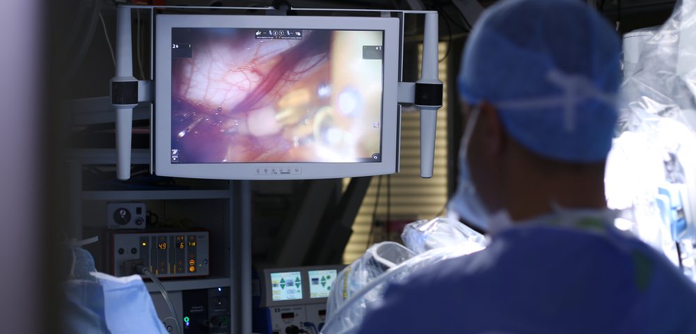
A new imaging-guided surgical technique that makes ovarian cancer tumors glow under near-infrared light can help surgeons remove tumors as small as 0.3 millimeters, a study in mice shows.
The approach also increased mice’s lifespan by 40% compared to mice treated with standard non-imaging-guided surgery. Researchers are now working to move this surgical technique into human trials.
The study, “Real-Time Single-Walled Carbon Nanotube-Based Fluorescence Imaging Improves Survival After Debulking Surgery In An Ovarian Cancer Model,” was published in the journal ACS Nano.
Ovarian cancer is among the deadliest cancers in women, mostly because nearly 75% of patients are diagnosed at an advanced stage, where treatment approaches fall short.
The gold-standard treatment for ovarian cancer is surgery — called debulking surgery — which aims to remove all visible tumor. However, some ovarian tumors are very small or hidden, making surgeons unable to remove the tumor entirely.
Scientists have been working on imaging devices that could help surgeons identify very small tumors during surgery, and found that a special type of imaging based on near-infrared light could penetrate living tissues with high efficacy while providing high resolution.
This imaging system uses chemical probes — made of single-walled carbon nanotubes — that bind SPARC, a protein found in invasive ovarian cancer cells, and emit fluorescent light when illuminated by a laser.
Researchers hoped that by providing continuous near-infrared light into the abdomen, surgeons would be able to detect even very small ovarian cancer tumors.
“You can’t have a patient in a CT machine or an MRI machine and have the surgeon perform this surgical debulking procedure at the same time, and you can’t expose the patient to X-ray radiation for multiple hours of the long surgery. This optics-based imaging system allows us to do that in a safe manner,” Neelkanth Bardhan, an oncology fellow at the Koch Institute and one of the lead authors of the paper, said in a press release.
“What’s nice about this system is that it allows for real-time information about the size, depth, and distribution of tumors,” said Angela Belcher, the James Mason Crafts Professor of Biological Engineering and Materials Science at the Massachusetts Institute of Technology (MIT).
In this study, researchers at MIT, along with surgeons and oncologists at Massachusetts General Hospital (MGH), set out to assess whether they could use this technology to locate tumors during ovarian cancer surgery in a mouse model of the disease.
Initially, researchers implanted ovarian tumors in the abdominal cavity of mice and then used the imaging-guided system to remove them. They were able to locate and remove tumors as small as 0.3 millimeters.
This is a significant finding, as no other imaging system is currently able to locate tumors that small during surgery.
Mice that received the imaging-guided surgery had no detectable tumors 10 days after the procedure, while control mice had many residual tumors left. By three weeks after the surgery, many of the tumors had grown back in the mice that had undergone imaging-guided surgery, but they still lived 40% longer than control mice.
“These data support the potential use of this novel imaging system in the intraoperative setting for the optical detection of residual malignant tissue at the time of surgical staging, and/or cytoreductive surgery in ovarian cancer patients,” said Alessandro Santin, a professor of obstetrics and gynecology and clinical research program leader at the Yale University School of Medicine.
Based on this data, researchers are seeking approval from the U.S. Food and Drug Administration for a Phase 1 clinical trial testing the imaging system in humans.
They also hope to adapt the system for monitoring patients at risk for tumor recurrence, and eventually for an early diagnosis of ovarian cancer.
“A major focus for us right now is developing the technology to be able to diagnose ovarian cancer early, in stage 1 or stage 2, before the disease becomes disseminated,” Belcher said. “That could have a huge impact on survival rates, because survival is related to the stage of detection.”

